I sistemi High-End sono sviluppati per rendere al massimo in ogni ambito diagnostico, per questo motivo ogni elemento che li compone corrisponde alla massima eccellenza tecnologica.
Costruiti con materiali d’avanguardia esprimono la più alta qualità diagnostica grazie a sofisticati algoritmi di processazione.
La grande potenza di calcolo consente l’utilizzo di trasduttori ad altissima densità di elementi per una “texture” estremamente raffinata dei dettagli anatomici.
L’elaborazione di immagini di nuova generazione permette ecografie 2D, 3D/4D ad alta risoluzione in tutti i campi di applicazione con il più alto livello diagnostico ed un sensazionale effetto immersivo.
Oltre la qualità ecografica su base tradizionale per lo studio della morfologia di organo su base qualitativa, questi sistemi consentono l’applicazione di softwares specifici dedicati a tutti i distretti corporei per aggiungere informazioni quantitativa e funzionale.
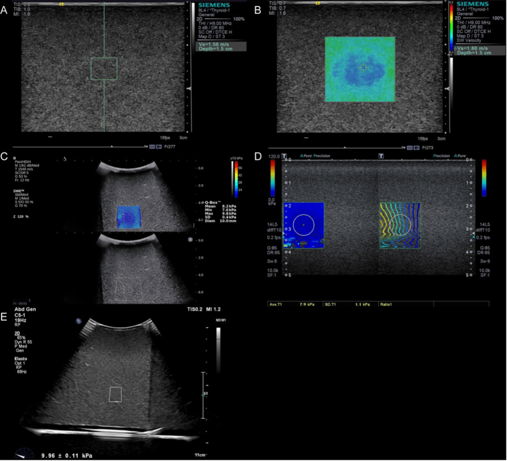
Per decenni, la biopsia epatica è stata il metodo convenzionale per la valutazione della malattia del fegato, più recentemente grazie alle moderne innovazioni la funzione elastosonografica ha introdotto una soluzione completa nella valutazione del trattamento e del monitoraggio delle epatopatie.
La funzione elastosonografica infatti fornisce una valutazione in tempo reale 2D della rigidità del parenchima epatico con la tecnologia 2D shear-wave (2D-SWE), è facile da eseguire, non è invasiva e offre la possibilità di effettuare misurazioni con parametri riproducibili per il monitoraggio della steatosi.
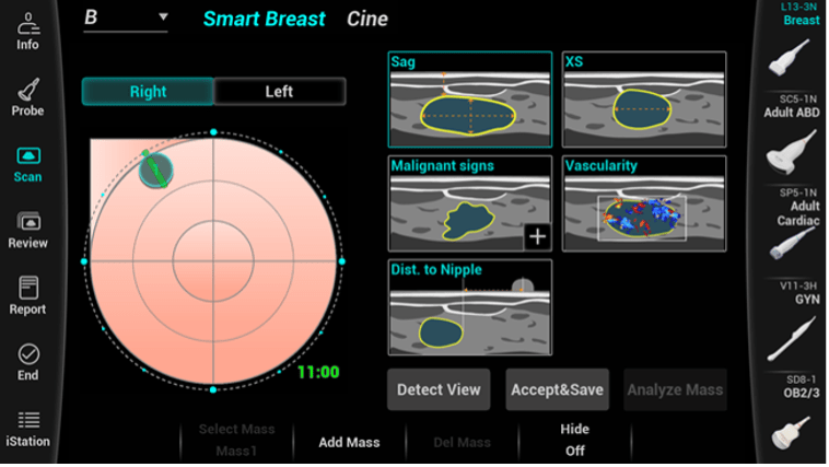
Le piattaforme ecografiche di nuova generazione assistono l’operatore con algoritmi di intelligenza artificiale anche nello studio della mammella.
Questi sistemi consentono una più completa valutazione del tessuto ghiandolare e delle lesioni mammarie attraverso funzioni di “lesion detection” che ne consentono la caratterizzazione del tessuto determinandone automaticamente la morfologia e grado di vascolarizzazione.
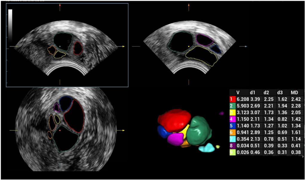
Un ulteriore avanzamento tecnologico è ottenuto attraverso l’utilizzo di sonde volumetriche 3D/4D, in grado di ottenere immagini sezioni volumetriche sulle quali è poi possibile una navigazione stratigrafica sui 3 piani spaziali X, Y, X.
Questa acquisizione permette si ottenere un volume costituito non più da pixel ( bidimensionali ) ma da voxcel (3D) ovvero ottenendo in vantaggio della dimensione spaziale delle strutture portando vantaggio nell’esecuzioni di alcuni distretti ecografici, come ad esempio l’utero e l’ovaio.
Ad esempio l’acquisizione automatizzata 3D nel monitoraggio follicolare permette di riconoscere e classificare follicoli per dimensione volume migliorando il confort della paziente riducendo drasticamente il tempo d’esame.
Anche in questo campo di applicazione le moderne tecnologie offrono diversi vantaggi diagnostici.
Nuovi algoritmi disponibili contribuiscono al completamento delle indagini ecografiche permettendo all’operatore di raccogliere informazioni precise sia morfologiche che emodinamiche.
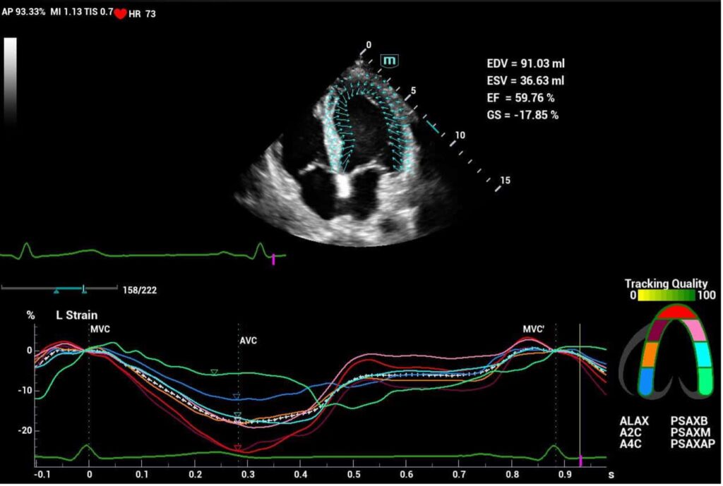
Nuovi software nell’ambito di studio ecocardiografico consentono di standardizzare l’esecuzione nella valutazione della cinetica parietale segmentaria con un alto grado predittivo sullo stato di salute dell’organo.